Bridging the Gap

Visual Tools for 3-Tesla MRI
Interpretation of the Deltoid Ligament

Project Summary

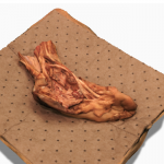
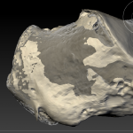
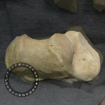
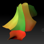
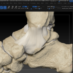
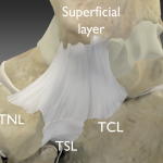
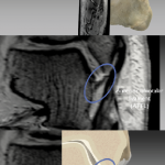
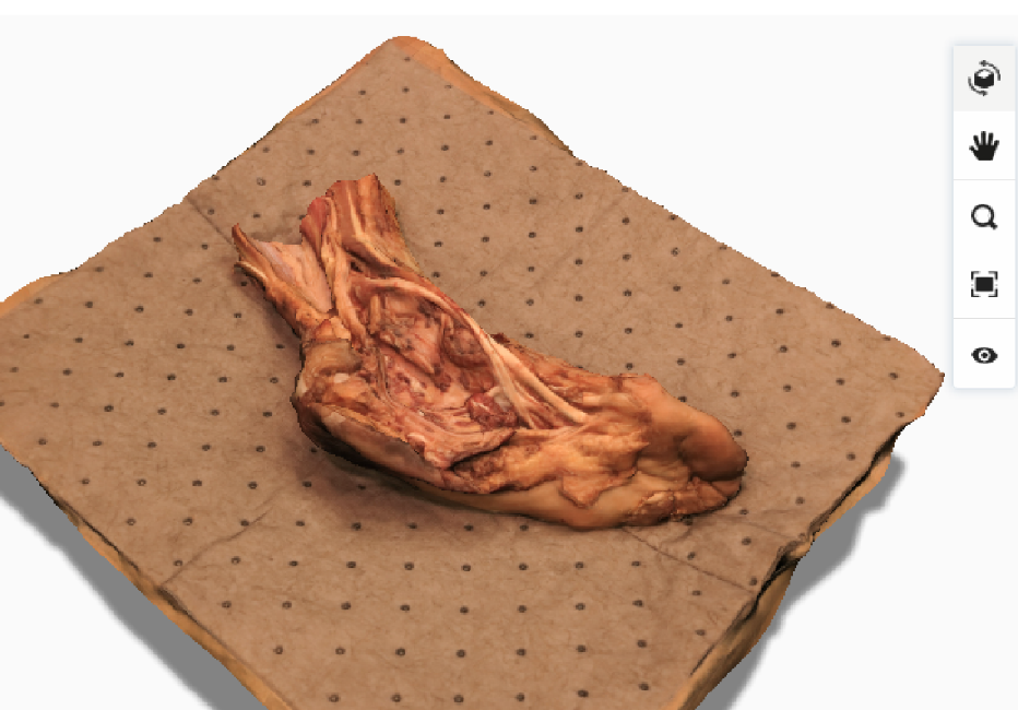
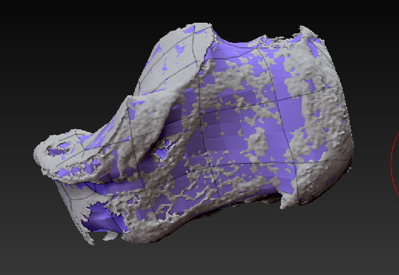
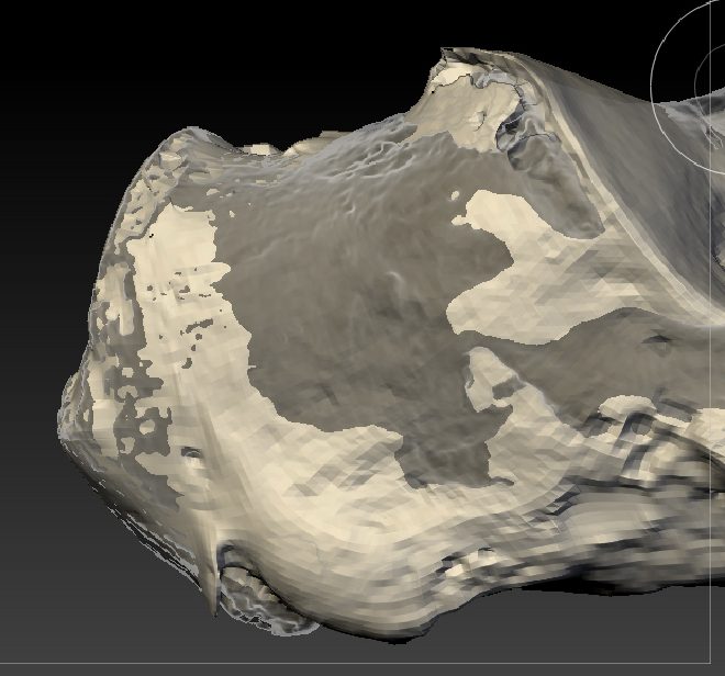
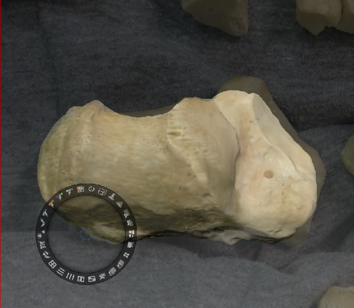
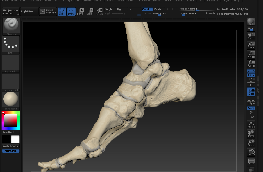
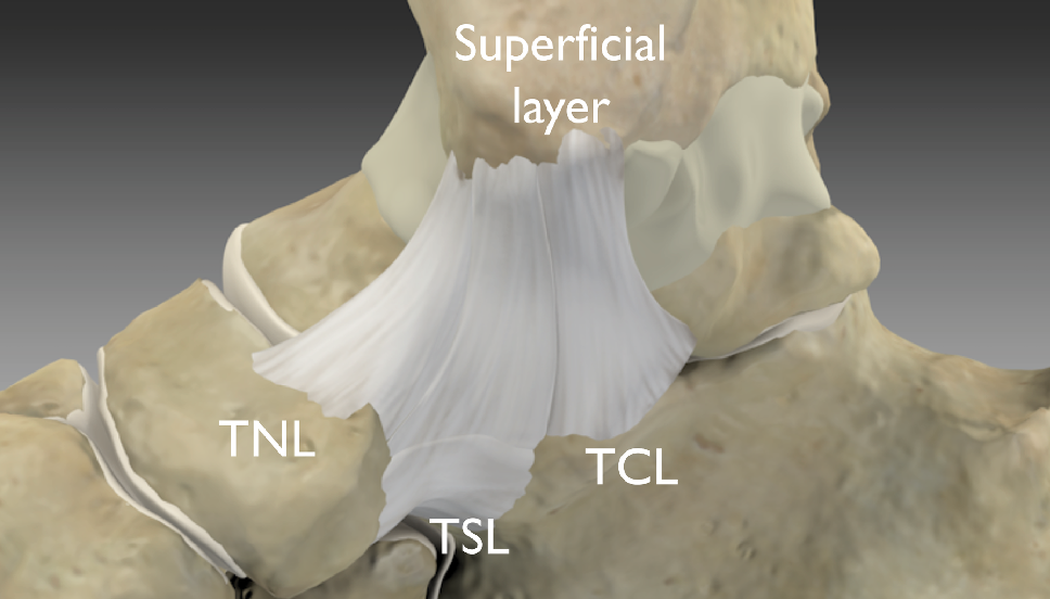
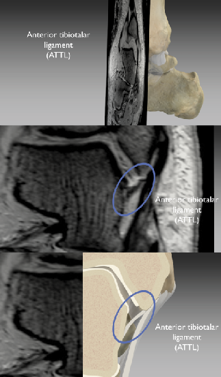
This thesis project involved the creation of detailed visual tools aimed to improve radiological residents’ anatomical understanding and high-field 3-Tesla MR image interpretation of the deltoid ligament (a.k.a. medial collateral ligament complex) of the ankle.  Photogrammetry models were created from the dissected specimens allowing interactive observation of the ankle dissections, and providing a spatial understanding of the deltoid ligament and surrounding structures. These models represent the specimens fairly accurately and provide a decent, though not ideal, substitute for individuals unable to view the specimens in person. A digital 3D model was produced utilizing CT, MR, surface laser scan, and photographic data. Created in ZBrush, it clearly shows the overall structure, layers, and ligamentous components of the deltoid ligament. This model was then utilized in an animation to demonstrate the anatomy and MR appearance of the deltoid ligament complex. The animation consists of three sections, which demonstrate the component anatomy, MR imaging characteristics, and injuries of the deltoid ligament. A narration guides the viewer through the animation and supplements the visual communications. Additionally, several novel and effective workflows were developed for this project that may prove useful in future medical visualization projects.

MR imaging is an effective method for evaluating the complex ligaments of the ankle; however, the accuracy of MR imaging diagnosis is heavily dependent on the training resources available to orthopedic radiology residents. Due to its complexity, much confusion surrounds the anatomical structure and MR diagnoses of the deltoid ligament components. The deltoid ligament consists of five components residing in two layers: a deep layer and a superficial layer. The deep layer consists of the anterior tibiotalar (ATTL) and posterior tibiotalar ligament (PTTL), both of which are intra-articular and lie within the synovial capsule of the talocrural joint. The superficial layer consists of the tibionavicular ligament (TNL), tibiospring ligament (TSL), and tibio calcaneal ligament (TCL), which lie superficial to the synovial capsule.

  • Create 3D digital dissection models using photogrammetry
  • Create a highly accurate digital model of the deltoid ligament
  • Create a 3D animation that demonstrates the components of the deltoid ligament and their attachments, correlates high-field MR images to the 3D structures of the anatomy, and illustrates the ligament injury grading/classification system.
  • Develop innovative approaches to combine data sets and software packages to create effective didactic visuals

Improvement in the abilities of orthopedic radiology residents to accurately:

  • Understand the structure of the deltoid ligament
  • Identify the components of the deltoid ligament on MR imaging
  • Distinguish normal variation from pathologies on MR imaging and identify grade/classification of injury.

Process

In order to fully understand and represent the deltoid ligament complex, this project was designed to have three main parts:

Ankle Dissection

Dissections were conducted and documented in order to more thoroughly understand the deltoid ligament’s structures and attachments.  Unpreserved specimens were MR and CT scanned prior to dissection. The deltoid ligament was exposed and the surrounding structures and spatial relationships were noted. The five components of the deltoid ligament were identified and the morphologies, attachment sites, and thicknesses of the components were observed.

Dissection Photogrammetry Models

Photogrammetry was used at different stages of dissection in order to capture digital dissection models. Photos were taken at set intervals using improved shooting techniques, processed in Adobe Bridge, and then uploaded to Autodesk 123D Catch to create 3D photogrammetry models of the dissection specimens. The raw photogrammetry models were cleaned up in ZBrush and imported into Maxon Cinema 4D to create turntables and interactive QTVR files. These models were used as reference while sculpting the ligaments for final animation model.

Imaging-based Model and Animation

A detailed digital model and animation were created using a combination of unpreserved cadaver CT and MR data, 3D surface laser scan data, and photos using the previous dissections and literature as references. Surface renders of bone from the ankle CT data were exported from OsiriX. ZBrush was utilized to create textured models of the bones and ligaments. A low-resolution animatic and the final 3D animation was created in Cinema 4D. Narration, labels, and titles in Adobe After Effects.

Innovations

The capabilities of 123D Catch, ZBrush, Cinema4D, and AfterEffects were combined in new ways throughout this project. The photogrammetry setup, shooting techniques, and processing were significantly improved beyond Autodesk guidelines. More widely applicable is the repair workflow used to create clean watertight meshes from suboptimal CT scan data which used a “digital putty” to fill in the damaged or missing areas of the scan data. Another innovation was the combined use of CT, MR, 3D laser scan, and photos together to create a highly accurate representation of the bones and ligaments. Additionally, a dynamic integration of MR imaging with a 3D moving cross section of the digital anatomical model was developed, allowed by utilizing MR and CT data from a single specimen.